Digital Dental X-Rays Raynham
Looking Beneath the Surface to Ensure Optimal Health

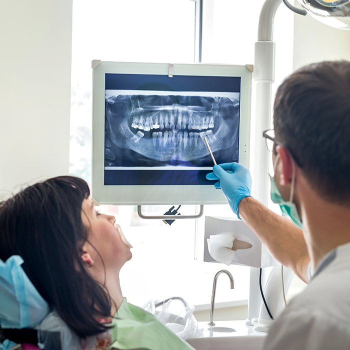
Dental digital X-rays are a modern and efficient way to capture detailed images of a patient’s teeth, gums, and bone structure using digital sensors instead of traditional film. These X-rays are commonly used in routine dental exams to detect issues like cavities, infections, bone loss, or impacted teeth, and to monitor overall oral health.
When Dr. Al and our team in Raynham take X-rays during your appointment, you can know with confidence that they’re putting your oral health’s best interests first. However, don’t hesitate to ask any questions about the process; we’re happy to show you just how beneficial these digital images are!
When Are Digital X-Rays Taken?
Dental X-rays serve an incredibly important role – simply put, the naked eye isn’t capable of seeing everything it needs to ensure optimal oral health. Dr. Al will normally take dental X-rays during the following situations:
- Routine Checkups: To monitor oral health and detect issues early.
- Suspected Tooth Decay or Cavities: When we need to look for cavities not visible during a visual examination.
- Infection or Abscess Detection: To spot infections below the surface of the gums or in the bone.
- Orthodontic Assessments: To assess the alignment of teeth and jaw structures, particularly for planning Invisalign or other treatments.
- Endodontic (Root Canal) Therapy: To evaluate the health of tooth roots.
Types of Dental Digital X-Rays
There are actually a few different types of X-rays used in dentistry, each with different purposes. However, all are useful. Here’s a closer look at some of the different X-rays Dr. Al might take when you come in for an appointment:
Intraoral X-Rays
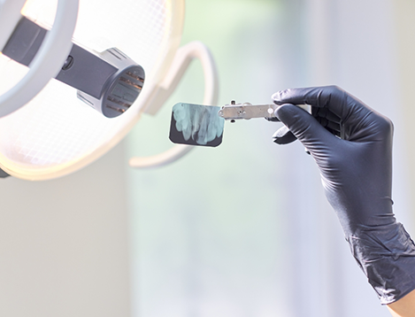
These are the most common type of dental X-ray. They involve placing a small sensor inside the mouth and include:
- Bitewing X-rays: Used to check for cavities between the teeth and assess the level of bone around them.
- Periapical X-rays: Show the entire tooth, from crown to root, and are useful for detecting issues beneath the gumline.
- Occlusal X-rays: Show the upper and lower jaw in one image to detect problems like cysts or fractures.
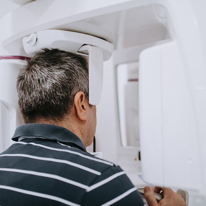
Extraoral X-Rays
These are taken outside the mouth and are used to capture images of the jaw, skull, and sinuses. Examples include:
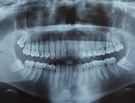
- Panoramic X-rays: Capture a wide view of the mouth, teeth, and jaw, useful for detecting issues with wisdom teeth, bone abnormalities, or jaw disorders.
- Cephalometric X-rays: Often used in orthodontics, these images help assess the relationship between the teeth and jaw.
The Safety & Benefits of Digital X-Rays

While X-rays do involve radiation exposure, the amount used in dental digital X-rays is minimal and considered safe for most patients. Dentists take precautions, such as using lead aprons and thyroid collars, to minimize exposure. When it comes to safety, digital X-rays typically use up to 80% less radiation than traditional film-based X-rays. Other key benefits include:
- Instant Image Availability: The images are available immediately after the X-ray is taken, allowing Dr. Al to quickly review and diagnose any issues.
- Higher Image Quality: Digital X-rays provide clearer, more detailed images that can be easily enhanced for better visualization of potential problems.
- Environmentally Friendly: Since digital X-rays don’t require the use of chemicals to develop images, they are more environmentally friendly compared to traditional X-ray film.
- Ease of Storage and Sharing: Digital images can be stored electronically and shared with specialists or other healthcare providers, making consultations and second opinions easier.
- Convenience: Since images can be accessed instantly and stored digitally, it’s easier for us to track a patient’s dental history over time.